Headache and dental occlusion, a possible correlation
9 February 2020
Adjusting occlusal appliance by functional evaluation of muscle activity
13 March 2020The case presented here was written by Dr Andrea Parenti, Dentist, Expert in Oral Surgery and Implant Prosthetics (1).
Presentation of the clinical case
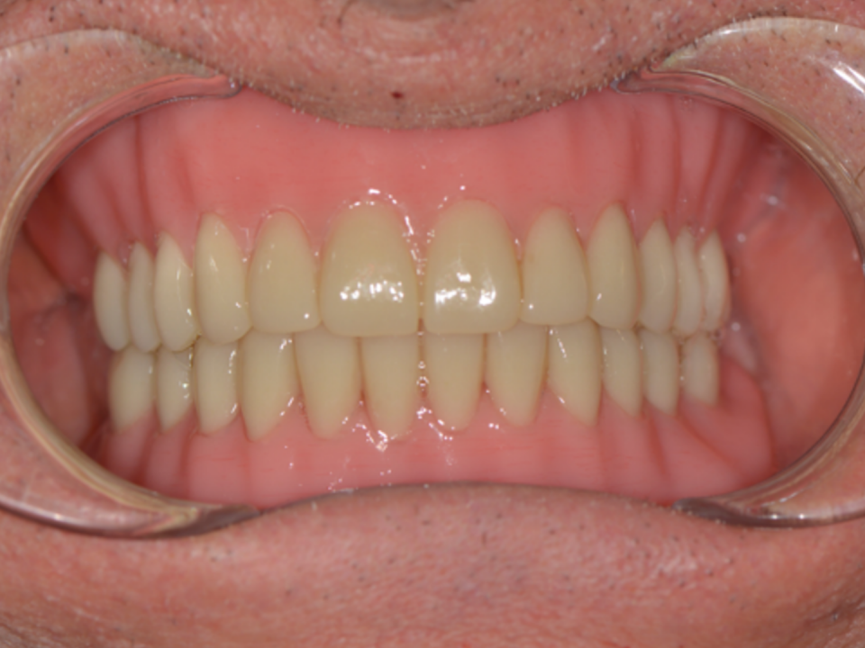
The clinical case discussed here shows a 51-year-old woman undergoing an implant-supported prosthetic rehabilitation of both upper and lower arches.The patient came to our observation after several years have passed since the last dental check-up.
His mouth presented various problems ranging from the absence of numerous dental elements, to the mobility of others due to the resorption of bone and gum around the same teeth. The teeth that did not show mobility in any case showed the same signs of resorption. All of this caused both functional and aesthetic deficits in our patient. During the first visit, we analyzed the patient’s general health situation and focused on his complaints and expectations. During the interview we established that the best solution for the patient was that of a complete fixed aesthetic and functional rehabilitation.


Figures 1-3: Initial situation – Pre-therapy


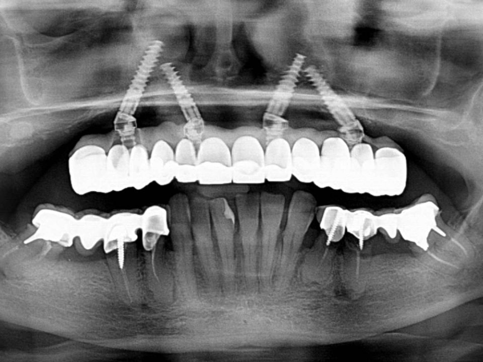
Figures 4-5: Initial situation – Pre-therapy (X-Ray and Teethan exam)
For this reason, specific radiological examinations were prescribed that allowed to proceed with a digital project and a simulation of the established result.
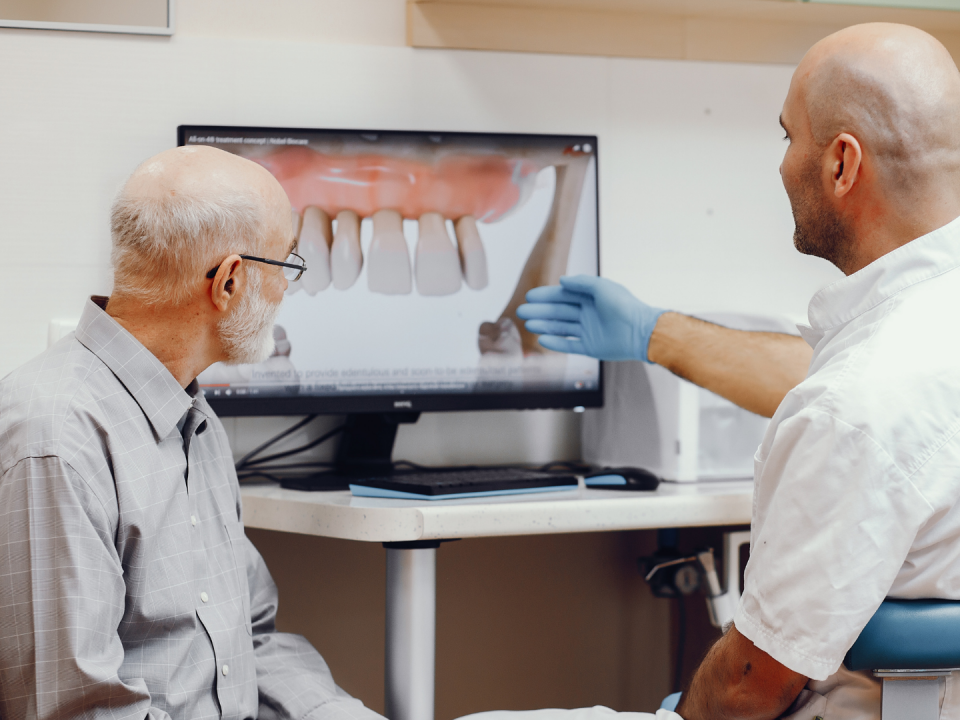
The figures show the simulation of the implant position and the simulation of the prosthetic rehabilitation that satisfies all the aesthetic and functional parameters required by the patient.
The figures show the simulation of the implant position and the simulation of the prosthetic rehabilitation that satisfies all the aesthetic and functional parameters required by the patient.


Figures 6-8: Planning
Therapy
The operative phase began with the upper surgery performed in conscious sedation to minimize the patient’s discomfort. In this way we proceeded to extract all the teeth of the upper arch, then we inserted the implants in the position corresponding to the position of the digital design and took a first impression.

Figure 9-10: Upper surgery


Figures 11-12: Lower surgery
The dental technician was able to create a fixed provisional screwed to the implants in 48 hours, which allowed the patient to regain much of the aesthetics and functionality lost over the years.
Obviously the provisional has some deficits when compared to the future definitive rehabilitation, but what matters is that in 2 days our patient has regained about 70% of what she had lost over time. All without complaining of discomfort either during surgery thanks to conscious sedation, or in the healing phase thanks to specific and targeted drug therapy.
After about 2 months we replicated the same approach to the lower arch. From this moment we and our patient only have to wait for the biological healing period, having a stable and respectable aesthetic and functional situation that has greatly improved the starting situation and which already has all the technical and functional characteristics to be included in the definitive rehabilitations.
In fact, by performing a digital instrumental examination of the occlusion thanks to the Teethan, it is possible to check the balance of the patient’s muscular work, which is optimal. This proves the correctness of the provisional rehabilitation. If we had not found positive values, the Teethan would have guided us in identifying the corrections to be made and subsequently in the quality control of these corrections.


Figures 13-14-15: Immediate load provisional rehabilitation


Figures 16-17: X-Ray and teethan exam
After about 5 months, the preparation phase of the definitive metal-free fixed restorations in zirconia and ceramic begins. These rehabilitations must fully reflect the aesthetic and functional parameters already included in the provisional restorations. Also in this phase the instrumental examinations such as intraoral radiographs, intra and extraoral photos and Teethan examination allow us to check precision, functionality and aesthetic performance of our products. After a series of 4 appointments, we can deliver the definitive rehabilitations and declare the rehabilitation of the patient completed, who solved all the deficits she had in less than 10 months from the first visit and which had prompted her to request the first appointment at our office.
In this clinical case Teethan allowed a check of the parameters correctness of both the provisional with immediate loading and fixed rehabilitation in monolithic zirconia. This was expressed in an excellent neuromuscular balance both during the treatment and at the end.
In this clinical case Teethan allowed a check of the parameters correctness of both the provisional with immediate loading and fixed rehabilitation in monolithic zirconia. This was expressed in an excellent neuromuscular balance both during the treatment and at the end.


Figures 18-19-20: Definitive rehabilitation


Figures 21-22: X-Ray and Teethan exam


Figures 23-24-25: Definitive rehabilitation
Curriculum Vitae Dott. Andrea Parenti
Graduated in Dentistry and Dental Prosthetics, Expert in Oral Surgery and Implant Prosthetics, since 2001 collaborator of Dr. T. Testori, Head of the Implantology and Oral Rehabilitation Department of the Dental Clinic (Director: Prof. RLWeinstein), University of Milan, IRCCS Galeazzi Orthopedic Institute, Milan.Since 2005 Tutor of the Implantology and Oral Rehabilitation Department of the same Institute Active Member SIO (Italian Society of Osseointegrated Implantology), AO (Academy of Osseointegration), EAO (Europen Academy of Osseointegration), SICOI (Italian Society of Oral Surgery and Implantology).
Member of the Active Membership Acceptance Commission of SICOI (Italian Society of Oral Surgery and Implantology) for the two-year period 2015-2016.
Founding Member A.I.S.G. (Advanced Implantology Study Group) and SISBO (Italian Society for the Study of Bisphosphonates in Odontostomatology). Speaker at National Courses and Congresses of Scientific Societies in the implant-prosthetic field.
Author of over 100 publications in national and international journals. Co-author of the book “Sinus surgery and therapeutic alternatives” Dr. T. Testori, Prof. R. L. Weinstein, Prof. S. Wallace, ed. Acme 2005 and the English language reissue “Maxillary Sinus Surgery”, Quintessence Publishing 2009.

