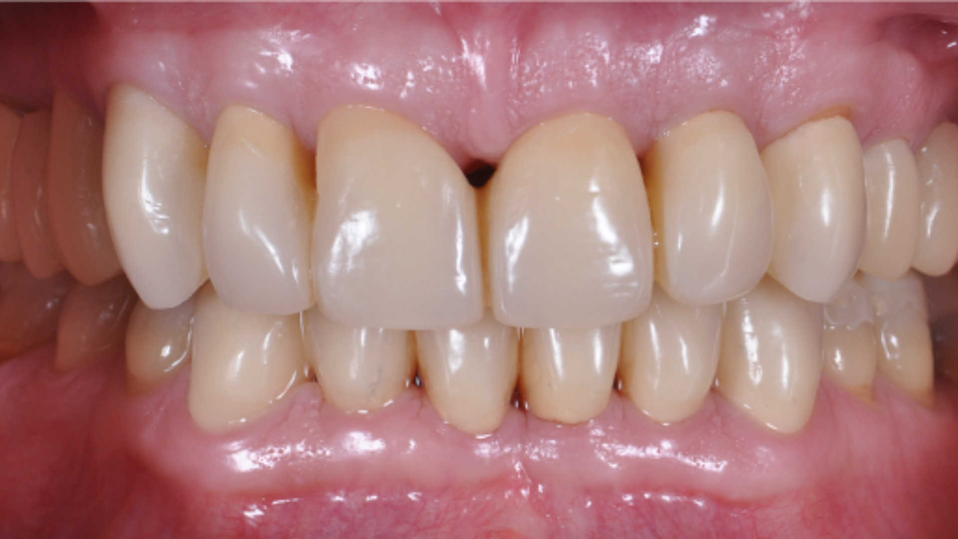
Complete rehabilitation with restoration of the vertical dimension of the lower third of the face
17 September 2021
Oral injuries in contact sports: the role of the mouthguard
15 October 2021The case presented here was elaborated by Dr. Filippo Cardarelli, Odontologist, specialized in Orthodontics.
Functional therapy should not be identified in the appliance or in the sequence of appliances that have the purpose of aligning the teeth, but in the use of a device that works in compliance with the physiology of the body with a functional goal. Skeletal and dental displacement always depend on neuromuscular function, for this reason the musculature must be taken into account for any type of evaluation. Elastodontic functional therapy fits perfectly within this scenario and aims to correct malocclusion and improve arch shape and craniofacial function. Within this therapy we find the AMCOP bioactivators, which consist of vestibular and lingual shields and are able to stimulate the muscles of chewing, lips and tongue. The therapeutic goal appears to be to eliminate dysfunctional counterforces by restoring the correct function of breathing, swallowing, lingual position, chewing, occlusion and posture. The proposed clinical case concerns a 10-year-old subject with skeletal class II, deep bite with 5 mm overjet and 6 mm overbite and shows how the elastodontic appliances are able to determine skeletal, dental and postural effects in a short time. The orthodontic movements were tested with surface electromyography (BTS TMJoint, Teethan).
10-year-old female patient with malocclusion. Skeletal class II deep bite with 5mm overjet and 6mm overbite, molar class II on the right and molar class I on the left. The arches show excess dento-maxillary disharmony with consequent crowding, gingival recession on element 41 (Figures 1a and 2a).
Premise
Functional therapy should not be identified in the appliance or in the sequence of appliances that have the purpose of aligning the teeth, but in the use of a device that works in compliance with the physiology of the body with a functional goal. Skeletal and dental displacement always depend on neuromuscular function, for this reason the musculature must be taken into account for any type of evaluation. Elastodontic functional therapy fits perfectly within this scenario and aims to correct malocclusion and improve arch shape and craniofacial function. Within this therapy we find the AMCOP bioactivators, which consist of vestibular and lingual shields and are able to stimulate the muscles of chewing, lips and tongue. The therapeutic goal appears to be to eliminate dysfunctional counterforces by restoring the correct function of breathing, swallowing, lingual position, chewing, occlusion and posture. The proposed clinical case concerns a 10-year-old subject with skeletal class II, deep bite with 5 mm overjet and 6 mm overbite and shows how the elastodontic appliances are able to determine skeletal, dental and postural effects in a short time. The orthodontic movements were tested with surface electromyography (BTS TMJoint, Teethan).
Materials and methods
10-year-old female patient with malocclusion. Skeletal class II deep bite with 5mm overjet and 6mm overbite, molar class II on the right and molar class I on the left. The arches show excess dento-maxillary disharmony with consequent crowding, gingival recession on element 41 (Figures 1a and 2a).


Photo 1a: Panoramic x-ray: beginning of therapy, 2010 / Photo 2a Intraoral frontal photograph: start of therapy, 2010
The subject also underwent latero-lateral teleradiography, an examination that revealed cervical hyperlordosis with hyperextension of the head on the neck, hyoid bone positioned low and back (Figure 3a). This position of the head in the sagittal plane forces an incorrect positioning of the Frankfurt plane to perform the teleradiography, since the patient is unable to maintain a correct head posture. The treatment plan provided for the use of only one elastodontic appliance worn for 2 hours during the day and every night for the first 6 months, and then only during the night. Once the molar ratio was corrected, the appliance was used as a restraint and to guide the exit of the last permanent teeth.

Photo 3a: Teleradiography: start of therapy, 2010
Results
After 24 months of elastodontic treatment a correct transverse dimension of the upper jaw was restored The physiological gingival level on element 41 has been completely restored. The teleradiography performed at the end of the treatment showed a normal cervical lordosis and a hyoid bone correctly positioned following correction of the skeletal class (Figure 3b) The achievement of normal occlusion was verified with the analysis of the masticatory muscles through electromyography (BTS TMJoint, Teethan). They were subjected to examination thanks to the use of 6 probes surface electomyography of the temporal, masseter and sternocleidomastoid muscles. The subject was asked to perform a test of maximum voluntary contraction on the cottons and subsequently a maximum contraction on the teeth. To test the activity of the sternocleidomastoids, a rotation of the head to the right and then to the left was performed, keeping the shoulder still. Electromyography was performed during therapy with and without an elastodontic appliance and highlighted the benefits it brings. In the test carried out with the device, compared to the test without, evaluating the parameters proposed by prof. Ferrario, an improvement in the POC (percentage overlap coefficients) for the temporal, masseter and sternocleidomastoid artery is shown; a posterior barycenter with values not far from the normal range, preferred over the anterior barycenter which could lead in the long term to an overload at the condylar level and could cause muscle-tension headaches (Figure 4).


Photo 3b: Teleradiography: end of therapy, 2013 / Figure 4: Results of the occlusal examination with BTS-TMjoint performed with and without an elastodontic appliance, during therapy
The result obtained from the examination confirms that the right muscle function has been restored, an indispensable requirement to ensure stability to the clinical case over time. The data obtained from the electromyographic examination is also very important for determining the resolution of functional malocclusion, linked to incorrect muscle functioning which in many cases orthodontics turns out to be the cause of the relapse.


Photo 1b: Panoramic x-ray: end of treatment, 2013 / Figure 2b: Intraoral frontal photographs: end of therapy, 2013
Discussion
The case in question was treated with elastodontic therapy and we can see how the therapy is natural, innovative, non-invasive and can be considered an extraordinary oro-craniofacial bio-orthopedics. Occlusal pathologies, and therefore malocclusions, are often a causal factor of many osteoarticular pathologies. Several studies show that skeletal class II is often associated with a posture advanced and hyperlordosis of the cervical spine, while class III is mostly associated with a backward posture. Through a careful analysis of the patient’s posture from the simple clinical examination to the latero-lateral x-ray teleradiography, a significant correlation between malocclusion and postural alterations is evident. With the achievement of normocclusion it was therefore also possible to correct the patient’s posture itself. In some cases, even physiotherapy or osteopathy sessions are useful to speed up and improve therapy In the last checkup, a further electromyographic test was performed, which shows the maintenance of the POC, the posterior barycenter and torsion (Figure 5).


Figure 5: Occlusal examination performed in the follow-up visit (2019) / Figure 3c: Frontal intraoral photographs: follow-up visit, 2019
Conclusion
The purpose of this work was to demonstrate the importance of early orthodontic treatment in order to simplify the treatment of malocclusions and reduce any recurrence. This method eliminates the need to resort to extractions, in harmony with the strict control of the anchoring and with the most conservative methods. The proposed therapies do not aim to align the teeth but to restore the occlusal function. Consequently, the restored occlusal function allows you to obtain the correct dental alignment and above all to preserve it over time. Preventive orthodontics using elastodontic devices therefore represents an important step forward in the field of orthodontics in developmental age since it is able to solve most orthodontic problems, transforming many of these cases into ideal occlusions from an aesthetic and functional and postural profile.
Resume
Degree in Dentistry and Dental Prosthetics from the University of L’Aquila, specialised in Orthodontics at the University of Milan. Since 2009, lecturer in Pediatric Dentistry at the University of Milan.Speaker in courses and congresses at national and international level. Creator of the orthodontic technique: Elastodontic Therapy®. Dr. Filippo Cardarelli deals exclusively with Orthodontics and Aesthetic Dentistry, applying innovative and minimally invasive diagnostic and therapeutic procedures, collaborating with other specialists in other disciplines to solve particularly complex cases.
Bibliography
- Besombes, Muzj et al. Contribution à l’ètude de la therapie fonction,nelle et de ses rèsultats. 34ème Congres de la socièt ODF 1961
- Bergersen EO. The performed retainer: principles and clinical applications. Am Assoc Orthod Audio-VisualLibrary, St Louis, 1970.
- Keski-Nisula K, Leo Keski-Nisula L, Saio H. et al. Changes after orthodontic intervention with eruption guidance appliance in the early mixed dentition. Angle Orthod 2008 Mar; 78(2):324-331.
- Janson GR, da Silva CC, Bergersen EO, et al. Eruption guidance appliance effects in the treatment of Class II, Division 1 malocclusions. Am J Orthod Dentofacial Orthop 2000 Feb;117(2):119-29.
- Keski-Nisula K, et al.: Orthodontic intervention in the early mixed dentition: A prospective, controlled study on the effects of the eruption guidance appliance. Am J Orthod Dentofacial Orthop; 2008;133, 2.
- De Farias Neto JP, de Santana JM, de Santana-Filho VJ, et al. Radiographic measurement of the cervical spine in patients with temporomandibular dysfunction. Arch Oral Biol 2010; 55 :670-678.
- Janson G, Nakamura A, Chiqueto K, et al. Treatment stability with the eruption guidance appliance. Am J Orthod Dentofacial Orthop 2007 Jun;131(6):717-28
- McNamara JA Jr, Howe RP, Dischinger TG. A comparison of the Herbst and Frankel appliances in the treatment of Class II malocclusions. Am J Orthod Dentofacial Orthop 1990;98: 134-144.
- Posen AL. The effect of premature loss of deciduous molars on premolar eruption. Angle Orthod 1965 Jul;35:249-52.
- Bergersen EO.:Preventive eruption guidance in the 5 to 7 year old. J. Clin. Orthod 1995;29 382-395.
- Baccetti T, Franchi L, Toth LR, Mc Namara JA JR. Treatment timing for Twin-block therapy. Am J Orthod 2003; 73:221-230.
- Mc Namara JA. Maxillary transverse deficiency. J Orthod Dentofacial Orthop 2000; 117:567 570.
- Wheeler TT, McGorray SP, Dolce C, et al. Effectiveness of early reatment of Class II malocclusion. Am J Orthod Dentofacial Orthop 2002; 121: 9-17.
- Sinclair PM, Little RM. Dentofacial maturation of untreated normals. Am J Orthod 1985 Aug; 88(2):146-56.
- Andrews LF. The straight wire appliance. Syllabus of philosophy and techniques. San Diego: Larry F. Andrews F. Foundation of Orthodontic Education and Research; 1975.
- Vanini L, D’Arcangelo C. Estetica, Funzione e Postura. Acme, Viterbo, 2018.
- Kendall F, Kendall McCreary E. I muscoli. Funzioni e test con postura e dolore. Verduci Editore, 1985.

