When the jaw clicks
26 October 2021
Parafunctions: bruxism and dental occlusion
30 October 2021This research paper, has been published in the 2020, November/December issue of the “Journal Of Biological Regulators&Homeostatic Agents” by Prof. Enrico Luigi Agliardi, together with Dr. Roberto Broggi, Dr. Matteo Clericò and Dr. Lavinia Sacchi.
JOURNAL OF BIOLOGICAL REGULATORS&HOMEOSTATIC AGENTS Vol. 34, no. 4 (S1), 77-0 (2020) All-on 4-type guided implant rehabilitation: a case report with proposed EMG protocol
Matteo Clericò (1), Roberto Broggi (2), Lavinia Sacchi (2), Enrico Agliardi (2,3)
(1) Doctor of Dental Medicine in Milan, Italy; (2) Department of Dentistry, IRCCS San Raffaele Hospital, Vita Salute University, Milan, Italy; (3) Chief of special rehabilitation surgery Department of dentistry S. Raffaele Hospital, Milan, Italy
Abstract
Rehabilitations with a low number of implants, which exploit the patient’s residual bone by tilting the fixtures, such as the All-on-Four® method, have now become a valid therapeutic option supported by an increasingly extensive literature on the subject, with very high success rates percentages. While the surgical protocol has now been validated for many years, the prosthetic and functional aspect is currently the subject of several developments relating mainly to the use of the chosen materials and the occlusal setup.
The objective of this article is to propose a prosthetic finalization protocol of a maxillary All-on-Four® with the aid of surface electromyography and the related analysis software (Teethan®), ensuring the delivery of a prosthesis with correct muscular function able to avoid functional overloads to the masticatory system and to the implants themselves.
Case Report
The all-on-4 implant rehabilitation method is widely described in the literature. The method allows to rehabilitate edentulous arches with implants and immediate fixed prosthesis. Malò et al report a survival rate of 95.4% after 7 years, with a good marginal bone level and a low complication rate. Most of the articles focus on survival rates.
However, few Authors describe the protocol exhaustively. Surface electromyography has proved to be an effective tool for assessing the correct functionality of the stomatognathic apparatus in relation to interarch dental relationships. Some Authors have created a clinical protocol for functional assessment using comparative values of muscle activity with and without dental contact; this protocol was inserted into a surface electromyography software to allow a quick and intuitive evaluation.
Other Authors have used magnetic resonance imaging to highlight the fit of the patient after implant rehabilitation. The objective of the presentation of this clinical case is to propose a protocol for guided EMG planning of an all-on-4 type rehabilitation, to ensure the delivery to the patient of a correct temporary and definitive prosthesis not only on the basis of the prosthetic parameters, but also functional ones. In fact, some Authors stress the presence of adequate occlusal support is a relevant factor in maintaining efficient chewing and suggest that it can play an indirect role in preventing the occurrence of temporomandibular dysfunction symptoms.
Case description (initial situation)
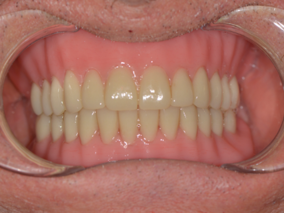
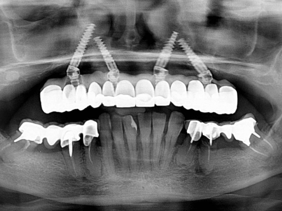
The 66-year-old patient comes to the Vita-salute San Raffaele hospital reporting pain and a history of recurrent abscesses in the upper arch. The clinical physical examination shows the loss of periodontal support (Fig. 1-4) as also confirmed by radiographic examinations (Fig. 5-8). The residual elements support a prosthetic rehabilitation from 16 to 23.
The patient reports a clinical history of chronic infections that undergo exacerbation with the formation of abscess collections treated with antibiotic therapy and with the appearance of pain on chewing. The antagonist arch also has a fixed implantprosthetic rehabilitation from 35 to 45 supported by residual dental elements. In consideration of the clinical situation, it was decided to intervene with the reclamation of the upper arch with the simultaneous insertion of 4 Osseo integrated implants according to the all-on-4 method.
Although the antagonist arch is not functional, it is left in the state in which it is, reserving, however, an evaluation before finalization to assess the need for any corrections of the occlusal plane.
A pre-surgery EMG (electromyography examination) recording is performed which however is not reliable due to the painful symptoms during maximum clamping.


Fig. 1-4. Pre-operative photographic records.


Fig. 5-8. First (intraoral) and second level (OPT) radiographic examinations that highlight the loss of periodontal support and the lack of congruity of the prosthetic rehabilitation.
Surgical phase
Before the surgery, the impressions of the two dental arches are taken and the provisional prosthetic product is packaged and delivered on the day of the surgery.
Once the patient has been accommodated according to safety protocols, a chlorhexidine rinse is performed. After conscious sedation, and plexus anesthesia, the reclamation of the upper arch is performed and the simultaneous insertion of 4 implants according to the all-on-4 protocol method.
The temporary prosthesis is delivered to the patient about 3 hours after surgery and functionalized with protected anterior contact as required by the original method (Fig. 9).
In this case, due to pain and sedation problems related to the surgery, the electromyographic examination is not performed immediately but at the first checkup after three weeks.

Fig. 9. Provisional prosthesis delivered on the day of surgery.
Check up at three weeks
Following tissue healing (Figures 10, 11) and an initial adaptation by the the patient to the new prosthetic product, the electromyographic evaluation is performed.
The examination showed an asymmetry of the contacts with an increase in the load on the left side arches and an anterior center of gravity; in accordance with the protocol which envisages reducing the load in the premolar area. After an initial post-surgical phase, the tissues must heal and seek new functionality, so it is necessary to optimize the occlusion to ensure correct distribution of the loads, balancing them (Fig. 12).


Fig. 10-11. Tissue healing 3 weeks after surgery.

Fig. 12. Electromyographic control recording at 3 weeks, left overload and anterior center of gravity were evident in accordance with the loading protocol of the all-on-4 method.
Three-month check-up
After the post-surgical checkup, occlusal balancing changes are made (Fig. 13). After 3 months, a control recording is made to verify functional adaptation. The electromyographic examination shows the anterior center of gravity with slight left overload (Fig. 14).

Fig. 13. Occlusal check at 3 months, following the protocol, full anterior and posterior skimming contacts are left.

Fig. 14. Three-month follow-up with slight left functional overload and anterior center of gravity.
Six-month check-up
Six months after surgery, tissue healing and osseointegration of the implants are now complete, so the work can be finalized.The impressions of the lower arch and the upper arch are taken. The finalization phase will include the delivery of a titanium meso-structure product (Procera REF), veneered in pink composite and individually cemented lithium disilicate teeth.
To optimize the process, a PMMA prototype of the final product is created which will serve not only as an aesthetic but also a functional try-in, allowing electromyographic recordings similar to the final one, in order to identify functional anomalies and correct them before moving on to the final one.
To correct the functional defects of the provisional, another electromyographic recording was performed (Fig. 15) with the aid of acetate shims, positioned between the arches (Fig. 16).
Thanks to a dimensional increase of the elements in the posterior sectors, the loads were redistributed to obtain more balanced recordings. The dental technician will thus be able to increase the vertical dimension, optimizing the supports.


Fig. 15-16. Electromyographic recording using acetate thicknesses to verify the functional adaptation to the posterior supports and the increase in the vertical dimension.
The new centric has been stabilized with silicone for occlusion and an electromyography is performed which confirms the desired occlusal balance (Fig. 17-18), everything is then transferred to the master model in the articulator to allow the dental technician to fill the inter-arch gap.
The measurement of the extent of the rise is carried out on the articulator and quantified in 1.5mm (Fig. 19-24).
The measurement of the extent of the rise is carried out on the articulator and quantified in 1.5mm (Fig. 19-24).


Fig. 17-18. New centric stabilized with silicone for occlusion and verified with EMG.


Fig. 19-24. Transfer of the new centric to the articulator.
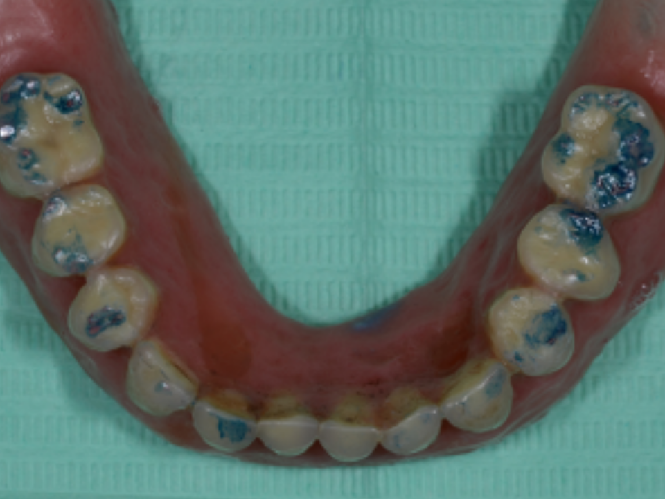
To obtain a correct occlusal plane, the programmed occlusal elevation was divided between the two arches and part of the occlusal elevation was performed in the lower arch on the patient’s old prosthetic artifact using a composite onlay (Fig. 25-30). In the upper arch, on the other hand, the temporary prosthesis and the master model are scanned and transferred to software. From this moment on, the design will be carried out on cad/cam software.
The sixths are then added, and the aesthetics modified according to the indications found in the photographs. Once the design phase is complete, the prototype in PMMA CAD / CAM is made and, after the cut back, pink resin is added to characterize the false gum.
The sixths are then added, and the aesthetics modified according to the indications found in the photographs. Once the design phase is complete, the prototype in PMMA CAD / CAM is made and, after the cut back, pink resin is added to characterize the false gum.


Fig. 25-30. Distribution of the occlusal elevation between the two arches.
After reassembling the upper arch and cementing the lower onlays, the control electromyography was re-performed with good functional parameters (Fig.31).

Fig. 31. Functional analysis of the prototype showing an almost total normalization of the parameters.
Although the upper arch is only a prototype, but equipped with sixths, the examination performed upon delivery can count on an adequate extension of the occlusal contacts that will be reproduced with the delivery of the final product which will have the current functionalized prosthesis as its basis.
The prototype is left in place for 7 days. A week later, in which the patient has tested the prosthesis, a new electromyographic trace is performed which shows improvement in muscle activity with parameters in the normal range. The functionalized prosthesis is returned to the laboratory for finalization in ceramic and will have to replicate the supports to maintain the adequate functionality detected and appreciated by the patient.
The electromyographic examination validated the correct centric, highlighting muscle activity in the normal range (Fig. 32).

Fig. 32. Normalization of functional parameters 7 days after delivery of the prototype.
The patient will be reviewed for the finalization of the therapy by converting the current upper prosthetic artifact into the definitive one by ceramizing the occlusal surfaces, always under electromyographic control.
The definitive prosthetic product is made using a CAD / CAM titanium structure whose structure is designed thanks to a cutback of the prototype itself, creating the retention abutments for each individual tooth. Following the production of the bar, a tryin is carried out with resin elements to verify its aesthetics and correct function, recording a new electromyographic trace, before the ceramic is made (Fig. 33-37).
Once the correct setting of the definitive has been confirmed, the finalization of the product is carried out; upon delivery, a new recording is made which highlights correct muscle function with reduction of torsion, capable of overloading the implant structures.
After 15 days, a new check shows the correct functionalization of the prosthesis as per initial programming (Fig. 38).


Fig. 33-37. Try-in with resin elements before finalizing the ceramic.

Fig. 38. Final control EMG in which the stability and normalization of the parameters is highlighted.
Discussion
From the results obtained, electromyography allows you to identify any imperfections in the occlusal load and correct them on the temporary restoration to obtain a correct and above all tolerated final rehabilitation by the patient.This statement is confirmed by the literature; some Authors point out that elderly patients accept implant rehabilitation with electromyography more easily, because they are more confident in bearing the definitive prosthesis.
However, other Authors point out that through an EMG examination the elderly with mainly mobile rehabilitations have greater loss of type II fibers at the periodontal level. Our study focuses on the design and optimization of the final prosthesis in an implant rehabilitation on a single case. Other Authors, on the other hand, have used electromyography to investigate the satisfaction of patients with implant rehabilitation, obtaining excellent results. The electromyography exam can be a valuable aid in implant-prosthetic rehabilitation to establish the right occlusal load; in fact, some Authors underline how the immediate implant load can influence a high crevicular fluid production.
Despite the limitations of our study due to the small sample and a non-randomized methodology, further studies are needed to deepen the subject on a higher sample of patients and a control group.
References
1. Agliardi E, Panigatti S, Clericò M, Villa C, Malò P. Immediate rehabilitation of the edentulous jaws with full fixed prostheses supported by four implants: interim results of a single cohort prospective study. Clin Oral Implants Res 2010; 21(5):459-65. doi: 10.1111/j.1600-0501.2009.01852.x.2. Capparé P, Teté G, Romanos GE, Nagni M, Sannino G, Gherlone EF. The ‘All-on-four’ protocol in HIVpositive patients: A prospective, longitudinal 7-year clinical study. Int J Oral Implantol (Berl) 2019; 12(4):501-510.
3. Agliardi EL, Tetè S, Romeo D, Malchiodi L, Gherlone E. Immediate function of partial fixed rehabilitation with axial and tilted implants having intrasinus insertion. J Craniofac Surg 2014; 25(3):851-5. doi: 10.1097/SCS.0000000000000959.
4. Maló P, de Araújo Nobre M, Lopes A, Ferro A, Botto J. The All-on-4 treatment concept for the rehabilitation of the completely edentulous mandible: A longitudinal study with 10 to 18 years of follow up. Clin Implant Dent Relat Res 2019; 21(4):565- 577. doi: 10.1111/cid.12769.
5. De Rossi M, Santos CM, Migliorança R, Regalo SC. All on Four® fixed implant support rehabilitation: a masticatory function study. Clin Implant Dent Relat Res 2014; 16(4):594-600. doi: 10.1111/cid.12031.
6. Bersani E, Regalo SC, Siéssere S, Santos CM, Chimello DT, De Oliveira RH, Semprini M. Implant supported prosthesis following Brånemark protocol on electromyography of masticatory muscles. J Oral Rehabil 2011; 38(9):668-73. doi: 10.1111/j.1365-2842.2011.02201. x.
7. Cattoni F, Tetè G, Uccioli R, Manazza F, Gastaldi G, Perani D. An fMRI Study on Self-Perception of Patients after Aesthetic Implant-Prosthetic Rehabilitation. Int J Environ Res Public Health 2020; 17(2):588. doi: 10.3390/ijerph17020588.
8. Ciancaglini R, Gherlone EF, Radaelli G. Association between loss of occlusal support and symptoms of functional disturbances of the masticatory system. J Oral Rehabil 1999; 26(3):248-53. doi: 10.1046/j.1365-2842.1999.00368.x.
9. Calderini A, Pantaleo G, Rossi A, Gazzolo D, Polizzi E. Adjunctive effect of chlorhexidine antiseptics in mechanical periodontal treatment: first results of a preliminary case series. Int J Dent Hyg 2013; 11(3):180-5. doi: 10.1111/idh.12009.
10. Polizzi E, Tetè G, Bova F, Pantaleo G, Gastaldi G, Capparè P, Gherlone E. Antibacterial properties and side effects of chlorhexidine based mouthwashes. A prospective, randomized clinical study. Journal of Osseointegration 2020; 12(1):230.
11. Wood KC, Berzins DW, Luo Q, Thompson GA, Toth JM, Nagy WW. Resistance to fracture of two all-ceramic crown materials following endodontic access. J Prosthet Dent 2006; 95(1):33-41. doi: 10.1016/j.prosdent.2005.11.003.
12. Sônego MV, Goiato MC, Dos Santos DM. Electromyography evaluation of masseter and temporalis, bite force, and quality of life in elderly patients during the adaptation of mandibular implant- supported overdentures. Clin Oral Implants Res 2017; 28(10):e169-e174. doi: 10.1111/clr.12980.
13. Guzmán-Venegas RA, Palma FH, Biotti P JL, de la Rosa FJB. Spectral components in electromyograms from four regions of the human masseter, in natural dentate and edentulous subjects with removable prostheses and implants. Arch Oral Biol 2018; 90:130-7. doi: 10.1016/j.archoralbio.2018.03.010.
14. Alam MK, Rahman SA, Basri R, Sing Yi TT, Si-Jie JW, Saha S. Dental Implants – Perceiving Patients’ Satisfaction in Relation to Clinical and Electromyography Study on Implant Patients. PLoS One 2015; 10(10):e0140438. doi: 10.1371/journal.pone.0140438.
15. Pellicer-Chover H, Viña-Almunia J, Romero-Millán J, Peñarrocha-Oltra D, García-Mira B, Peñarrocha-Diago M. Influence of occlusal loading on periimplant clinical parameters. A pilot study. Med Oral Patol Oral Cir Bucal 2014; 19(3):e302-7. doi: 10.4317/medoral.19477.

